PLIN3 as a Regulator of Lipid-droplet Biogenesis
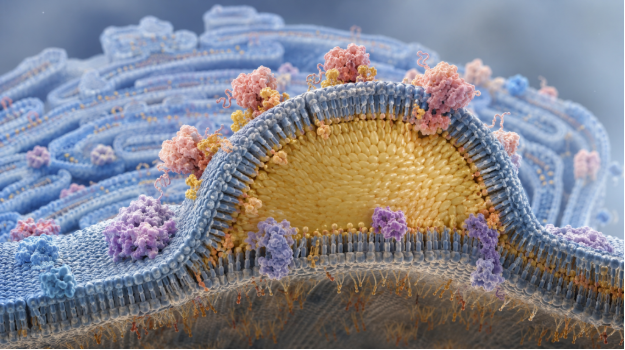
Disruption in the formation of lipid droplets alters energy balance, membrane homeostasis, stress responses, and disease-associated signaling. To understand the formation of these organelles, it is important to understand the proteins that recognize nascent droplet sites and organize the surrounding membrane.
Traditionally, researchers have used Perilipin 3 (also known as TIP47) as a marker of newly forming lipid droplets. However, recent structural and functional evidence suggests that Perilipin 3 (PLIN3) may:
- Sense lipid composition
- Remodel endoplasmic-reticulum (ER) membranes
- Recruit components needed for lipid-droplet biogenesis
PLIN3 Within the Perilipin Family
Lipid droplets consist of a neutral-lipid core (primarily triacylglycerols and sterol esters) which is surrounded by a phospholipid monolayer. Members of the perilipin family bind to this phospholipid monolayer and regulate how stored lipids are packaged, protected, and mobilized. PLIN3 is expressed widely and is recruited early in droplet development. It can move from the cytosol to ER sites where neutral lipids begin to accumulate.
PLIN3 contains an N-terminal Perilipin-ADRP-TIP47 (PAT) domain, which is a region of 11-mer repeats, and a C-terminal four-helix bundle. The PAT domain and 11-mer recognize lipid environments associated with emerging droplets. The contribution of the four-helix bundle appears to depend on membrane composition and experimental conditions.
Recruitment of PLIN3 to Nascent Lipid Droplets
Lipid-droplet biogenesis begins at specialized ER subdomains. Neutral lipids accumulate between the ER membrane leaflets and form a lipid lens that grows and eventually buds toward the cytosol. This process requires coordinated changes in lipid composition, membrane curvature, and protein localization.
PLIN3 is recruited during these early stages. Gaps in the membrane surface that expose hydrophobic regions to amphipathic protein segments influence the targeting of PLIN3.
PLIN3 can therefore recognize the physical properties of a developing droplet while also responding to particular lipids concentrated at the biogenesis site. This combined sensitivity may explain why PLIN3 prefers binding to nascent droplets rather than binding indiscriminately to cellular membranes.
Diacylglycerol As a Determinant of PLIN3 Localization
Diacylglycerol (DAG) serves both as a signaling lipid and a direct precursor of triacylglycerol. Evidence indicates that DAG becomes enriched in discrete ER domains where lipid droplets form.
In vitro studies show that PLIN3 binds liposomes that contain DAG.
Cell-based experiments link DAG enrichment to the formation of PLIN3-associated membrane domains.
Research published in Frontiers in Cell and Developmental Biology found that membrane-anchored perilipins induced expanded ER domains with lipid-droplet-like properties.
Enriched in DAG, these domains contained early biogenesis proteins, including seipin and Pex30. They could also develop into nascent droplets when neutral-lipid synthesis was induced.
These findings suggest that DAG-rich membranes recruit PLIN3. Then, PLIN3 helps organize membrane domains that concentrate the machinery required for droplet formation.
This shows that PLIN3 is more than just a surface marker. It may help establish or stabilize a local membrane environment from which a lipid droplet can emerge.
Structural Basis of PLIN3 Membrane Association
Much of the PAT domain and 11-mer repeat region is disordered in solution. When PLIN3 encounters an appropriate membrane, segments within these regions adopt ordered amphipathic alpha helices.
The hydrophobic faces of ordered amphipathic alpha helices can interact with membrane-packing defects, with their polar faces remaining exposed to the aqueous cytosol.
Work published in Nature Communications identified an expanded PAT domain that preferentially associates with DAG-enriched membranes. Membrane binding produced a disorder-to-order transition and a folded but dynamic structure. A related study in the Journal of Biological Chemistry identified conserved PAT-domain residues that mediate binding to membranes containing DAG.
Altering these residues weakened membrane association, providing evidence that PLIN3 recruitment depends on molecular determinants rather than nonspecific hydrophobic binding.
Conclusions from these findings support that PLIN3 behaves as a lipid-responsive membrane sensor. DAG accumulation and packing defects create a favorable binding surface. PLIN3 then undergoes conformational changes that stabilize its membrane-associated state.
PLIN3 as a Regulator—Not Merely a Marker
PLIN3 may do more than mark newly forming lipid droplets. It can bind DAG-enriched membranes and undergo conformational changes. It also associates with early lipid-droplet biogenesis proteins. This suggests that it may help organize the membrane environment required for droplet formation. Rather than simply arriving at a developing droplet, PLIN3 may help establish or stabilize the membrane domains from which droplets emerge. However, whether PLIN3 initiates these changes or stabilizes domains formed by other mechanisms remains to be determined.
Opportunities for PLIN3 Small-molecule Research
The emerging mechanism provides a foundation for PLIN3 small molecule research, meaning the discovery of compounds that modulate PLIN3 rather than treating PLIN3 itself as a small molecule. Potential compounds could:
- interfere with PAT-domain folding
- alter PLIN3 recognition of DAG-rich membranes
- stabilize a conformation that changes its residence time at nascent droplets
Liposome-binding assays could quantify compound-dependent changes in PLIN3 recruitment. Cellular imaging could measure droplet number, size, and PLIN3 localization. Structural assays could determine whether candidate compounds bind PLIN3 directly or merely alter membrane properties. That distinction is critical because a lipid-active compound might change PLIN3 localization indirectly without engaging the protein.
Conserved features across the perilipin family may make it difficult to modulate PLIN3 without affecting related proteins. Studies so far do not establish a validated PLIN3 inhibitor, activator, or therapeutic ligand. PLIN3 should therefore be described as an emerging experimental target.